


RESEARCH
1. Multifunctional nanoparticles based therapeutics for cancer imaging and therapy
Multifunctional nanoparticles have shown tremendous potential in the delivery of cancer drugs and imaging reagents for cancer therapy and imaging. Our lab focuses on the development of multifunctional nanoparticles from both biology and engineering perspectives. We have efficiently combined formulation design, micro/nanoengineering method with delivery mechanism study to design/produce multifunctional nanoparticles with high efficacy for cancer diagnosis and therapy.
1.1. Formulation design of multifunctional nanoparticles
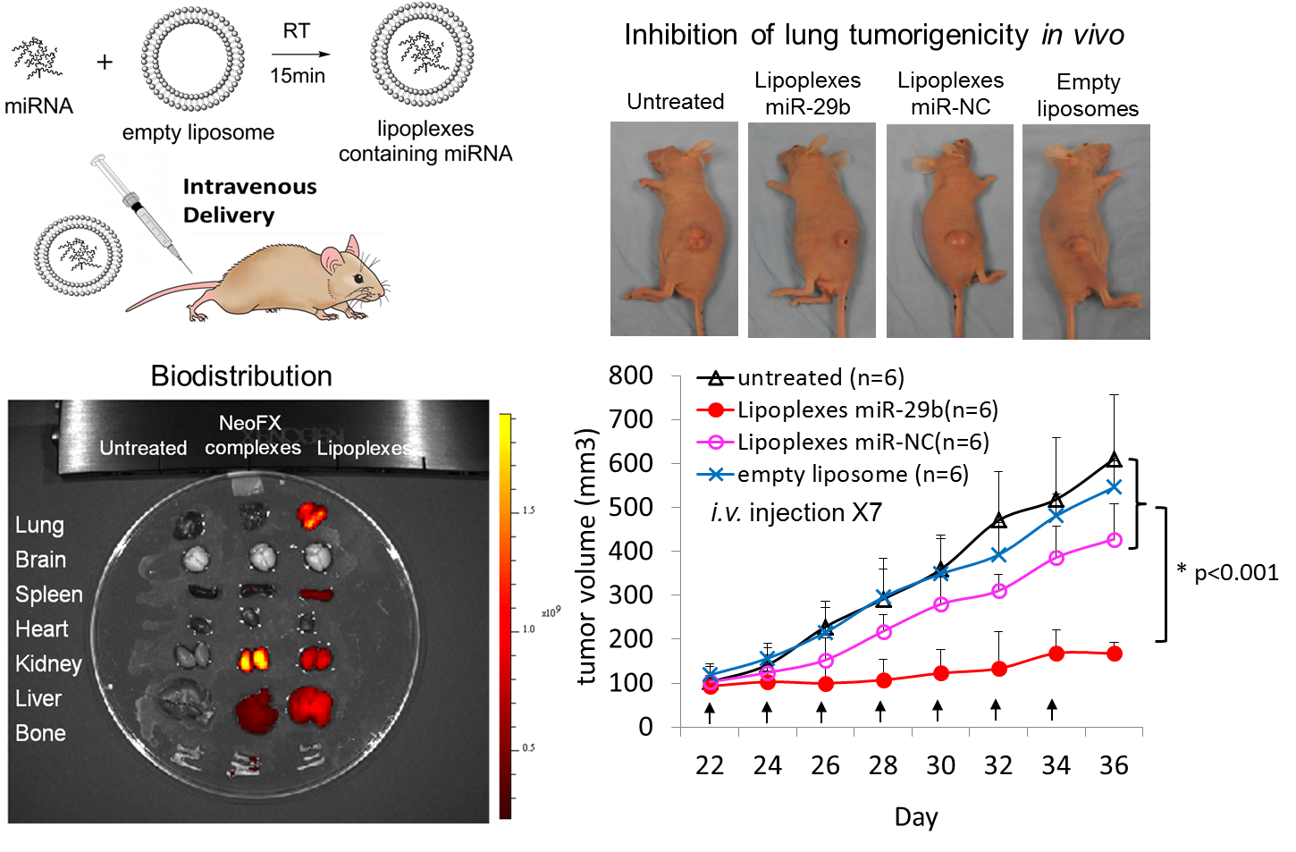
We have developed a series of multifunctional nanoparticles using lipids, polymers and inorganic materials as building blocks to deliver various drugs (chemotherapy drugs, microRNAs, siRNAs, DNAs and proteins) and contrast reagents for cancer therapy and imaging. Following example shows a cationic lipoplex nanoparticles delivery system that efficiently delivered miRNA-29b to tumor sites and inhibited the tumorigenicity of non-small cell lung cancer (NSCLC).
Multifunctional nanoparticles have shown tremendous potential in the delivery of cancer drugs and imaging reagents for cancer therapy and imaging. Our lab focuses on the development of multifunctional nanoparticles from both biology and engineering perspectives. We have efficiently combined formulation design, micro/nanoengineering method with delivery mechanism study to design/produce multifunctional nanoparticles with high efficacy for cancer diagnosis and therapy.
1.1. Formulation design of multifunctional nanoparticles
We have developed a series of multifunctional nanoparticles using lipids, polymers and inorganic materials as building blocks to deliver various drugs (chemotherapy drugs, microRNAs, siRNAs, DNAs and proteins) and contrast reagents for cancer therapy and imaging. Following example shows a cationic lipoplex nanoparticles delivery system that efficiently delivered miRNA-29b to tumor sites and inhibited the tumorigenicity of non-small cell lung cancer (NSCLC).
1.2. Micro/Nanoengineering method development for multifunctional nanoparticles production
2. In vitro diagnostics for cancer screening, early detection and prognosis
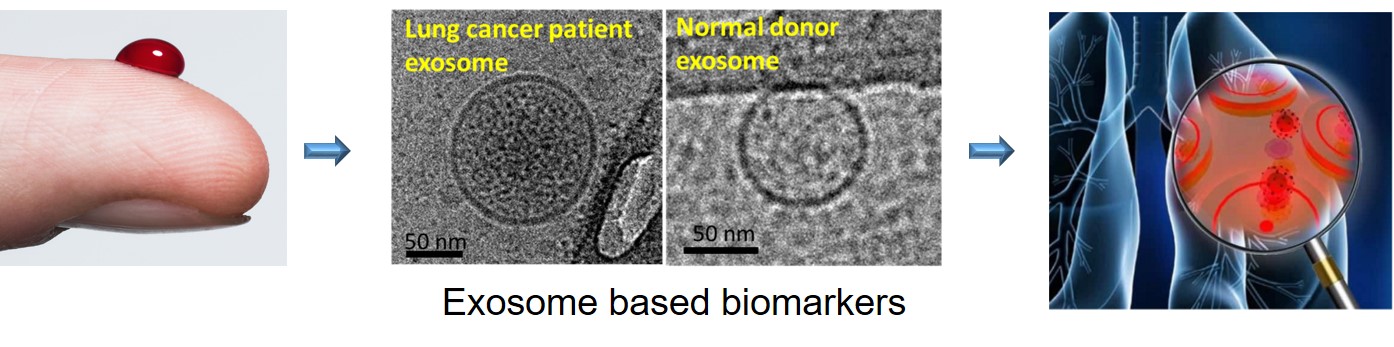
Exosomes are nano-sized vesicles released by all type of cells in the body. They transfer biomolecules, such as DNA fragments, microRNAs, mRNAs, proteins and lipids from parent cells to recipient cells for cell-to-cell communications. Exosomes, especially those released by cancer cells, play important roles in cancer development, angiogenesis, metastasis, immune response, and drug resistance. Therefore, exosomes are promising biomarkers in cancer screening, early detection, treatment response monitoring and prognosis.
We have developed sensitive, simple, fast and cost-effective in vitro diagnostic devices that capture tumor-derived exosomes and detect biomarkers carried by exosomes for cancer diagnosis. We have been actively collaborated with clinicians and scientists from Roswell Park Comprehensive Cancer Center and Mayo Clinic to evaluate the diagnostic performance of these devices in several types of cancer, such as lung cancer, breast cancer and colorectal cancer and brain cancer.
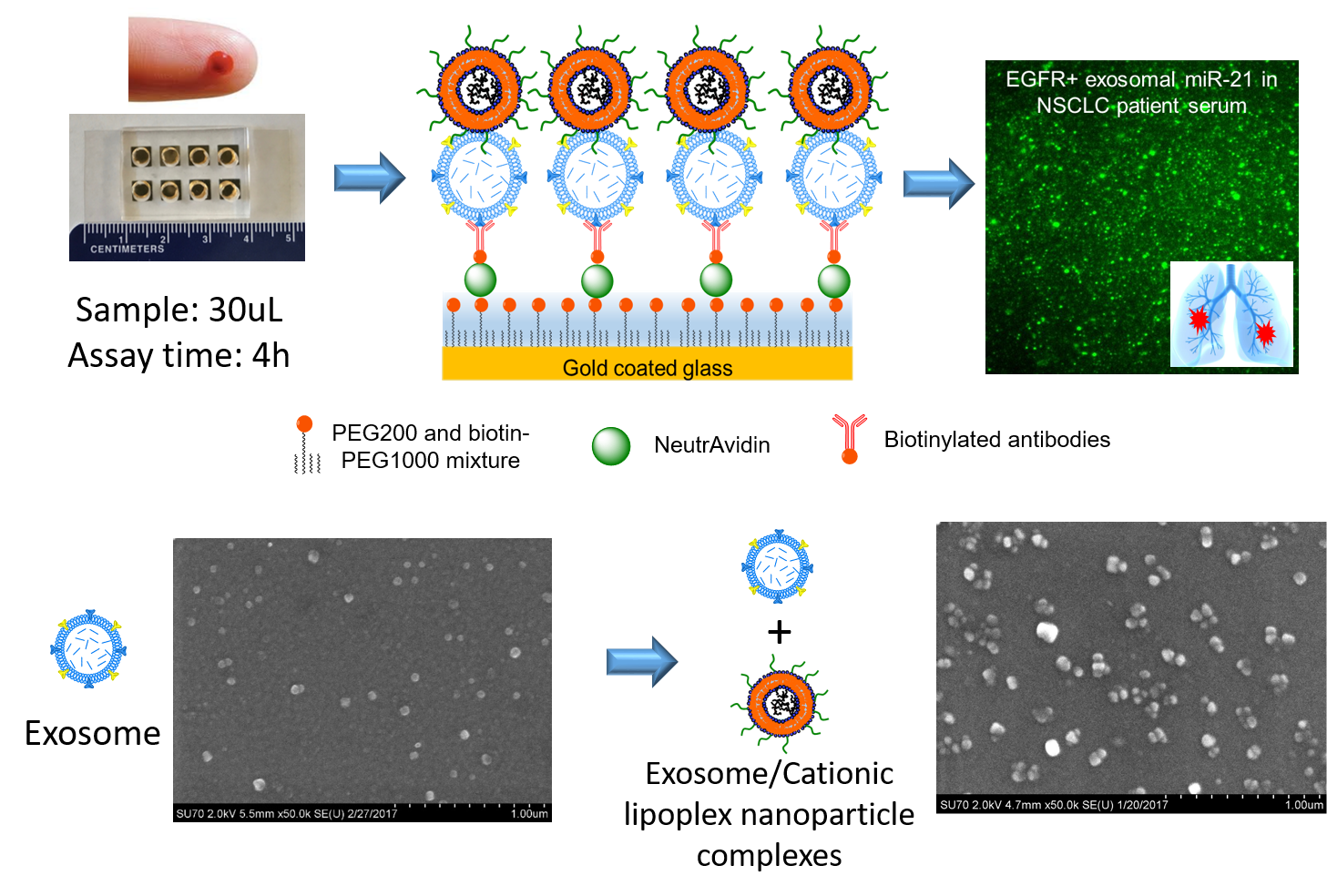
In the immuno-biochip, tumor-derived exosomes are captured by antibodies against tumor-associated proteins. Then cationic lipoplex nanoparticles containing sensing probes, such as molecular beacons are added and fuse with exosomes through electrostatic interaction to form CLN-exosome complexes. Inside CLN-exosome complexes, molecular beacons detect RNA targets and generate fluorescent signals, which are used to measure the expression of exosomal RNAs for cancer diagnosis. Compared with conventional technologies, our immuno-biochip provides higher detection sensitivity and specificity, consumes less serum samples (30 uL vs 100 uL), and shortens assay time (4h vs 24h).
1.2.1. Microfluidic preparation of multifunctional lipoplexes nanoparticles
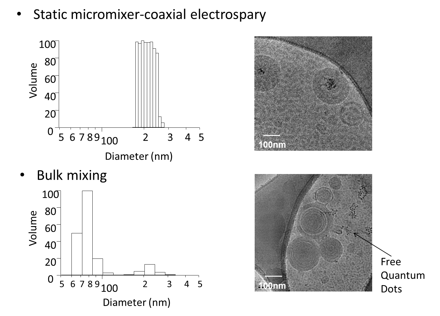
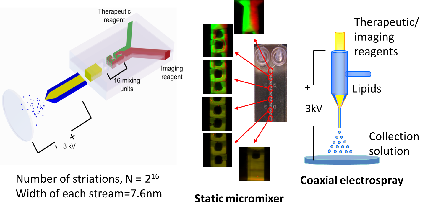
We have developed a novel technique, called the static micromixer-coaxial electrospray (MCE), to produce theranostic lipoplexes containing quantum dots (QD605 as the model imaging reagent ) and Cy5-labeled antisense oligodeoxynucleotides (Cy5-G3139 as therapeutic drug). MCE device is able to produce uniform theranostic lipoplexes in a single step with high reproducibility. Theranostic lipoplexes also showed good therapeutic and imaging ability with little toxicity. The MCE device will benefit the development of theranostic complexes and facilitate clinical transition.
We have developed a novel technique, called the static micromixer-coaxial electrospray (MCE), to produce theranostic lipoplexes containing quantum dots (QD605 as the model imaging reagent ) and Cy5-labeled antisense oligodeoxynucleotides (Cy5-G3139 as therapeutic drug). MCE device is able to produce uniform theranostic lipoplexes in a single step with high reproducibility. Theranostic lipoplexes also showed good therapeutic and imaging ability with little toxicity. The MCE device will benefit the development of theranostic complexes and facilitate clinical transition.
Production of multicomponent nanoparticles with uniform size, structure and composition in a controlled and reproducible process is critical for their success in clinical translation. Our lab has developed micro/ nanoengineering methods that offer better control over the nanoparticle formation compared to conventional methods.
1.2.2. Microwell array based production of multifunctional lipoplexes nanoparticles
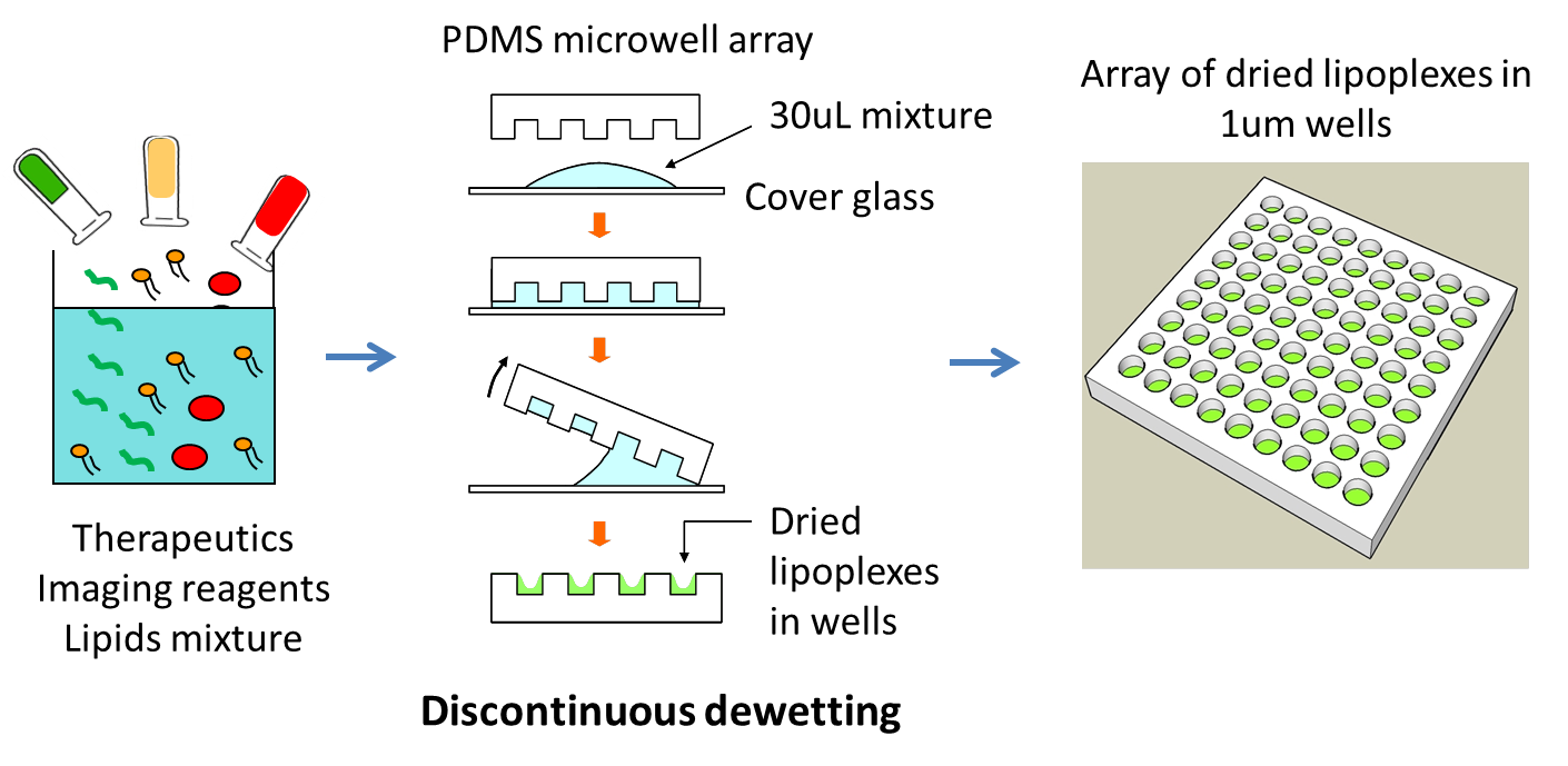
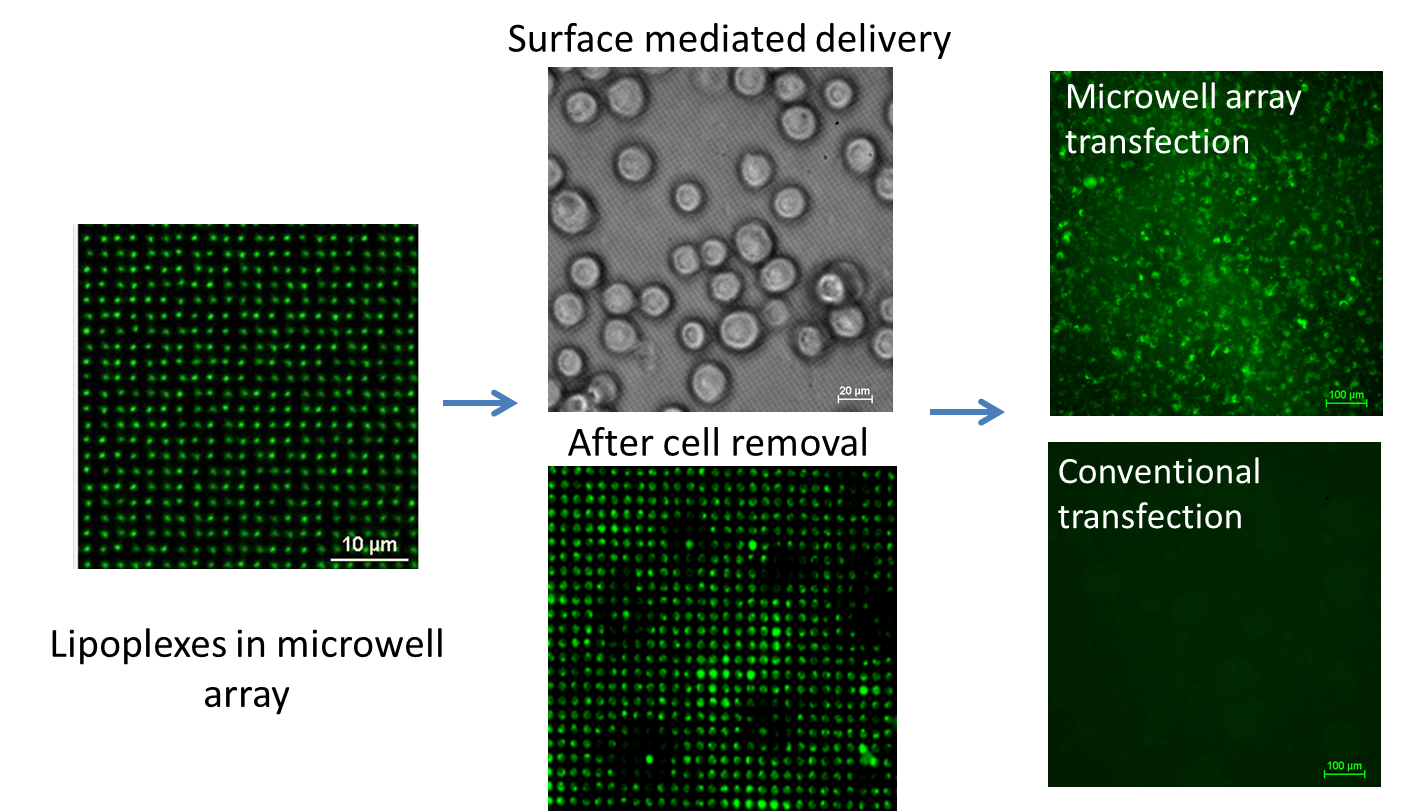
We prepared lipoplexes inside the microwell array by a discontinuous dewetting technique. This technique represents a straightforward way to quickly and uniformly fill very small wells with liquid by exploiting the differences in the interfacial free energies of the substrate and the liquid. Microwell array mediated delivery of lipoplexes containing nucleic acids is a novel, simple and cost-effective method. It showed much more efficient delivery than the conventional transfection, and thus significantly enhanced the therapeutic efficacy at much lower effective dose of therapeutic reagents.
We prepared lipoplexes inside the microwell array by a discontinuous dewetting technique. This technique represents a straightforward way to quickly and uniformly fill very small wells with liquid by exploiting the differences in the interfacial free energies of the substrate and the liquid. Microwell array mediated delivery of lipoplexes containing nucleic acids is a novel, simple and cost-effective method. It showed much more efficient delivery than the conventional transfection, and thus significantly enhanced the therapeutic efficacy at much lower effective dose of therapeutic reagents.
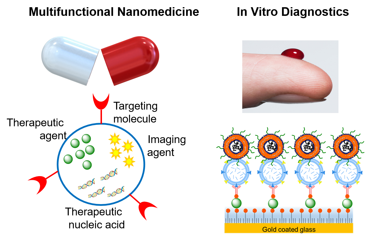
Our research focuses on: (1) developing multifunctional nanoparticles based nanomedicine as new and potent therapies for cancer and chronic diseases; and (2) developing innovative, nanotechnology-enabled biosensors that detect circulating biomarkers in body fluids (such as blood and urine) as non- or minimally-invasive in vitro diagnostics for cancer screening, early detection, treatment response monitoring and prognosis.
2.1. Immuno-biochip detects exosomal RNAs for cancer diagnosis
Immuno-biochip
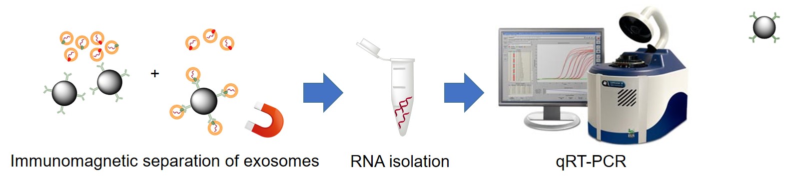
Conventional method
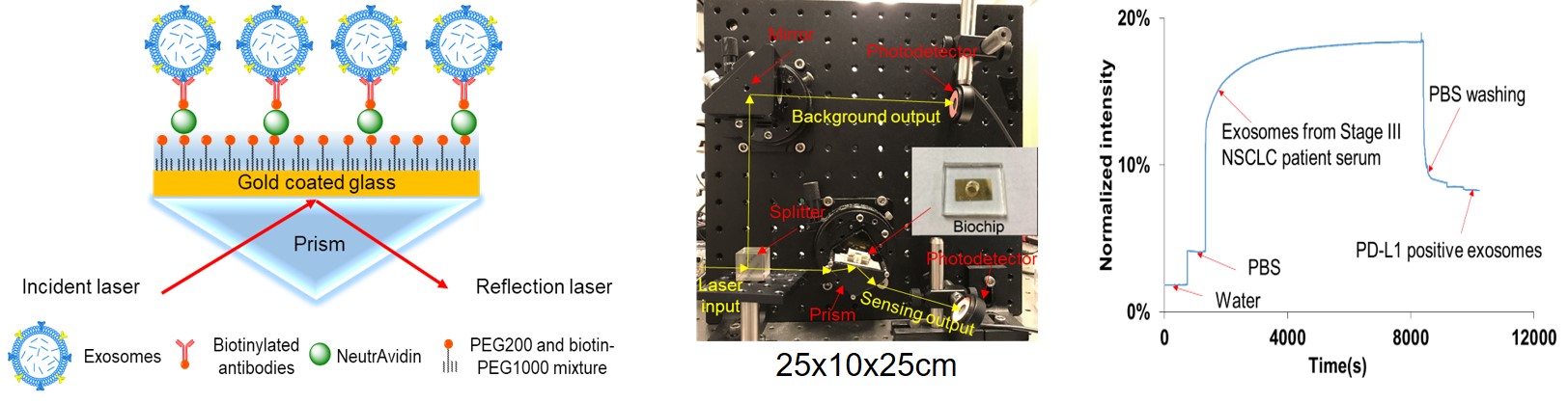
2.2. Compact surface plasmon resonance (SPR) biosensor detects exosomal proteins for cancer diagnosis
Among many detection techniques, surface plasmon resonance (SPR) is a highly sensitive, label-free and real-time optical detection method. Commercial prism-based wavelength/angular-modulated SPR sensors afford high sensitivity and resolution, but their large footprint and high cost limit their adaptability for clinical settings. We have developed an intensity-modulated, compact SPR biosensor (25 cm × 10 cm × 25 cm) that detects exosomal proteins for cancer diagnosis. We have demonstrated the feasibility of the compact SPR biosensor in lung cancer diagnosis using exosomal epidermal growth factor receptor (EGFR) and programmed death-ligand 1 (PD-L1) as biomarkers. The compact SPR biosensor showed higher detection sensitivity than ELISA and similar sensing accuracy as ELISA.

Another example is a MnO2 nanotubes based imaging system that can be used as a glutathione-activated agent for photoacoustic imaging and fluoresnece imaging. We have demonstretad its applicaitons in photoacoustic imaging of glutathione for melanma diagnosis and fluorescence detection of oncomicroRNAs (miR-21 and miR-155) in breast cancer diagnosis.
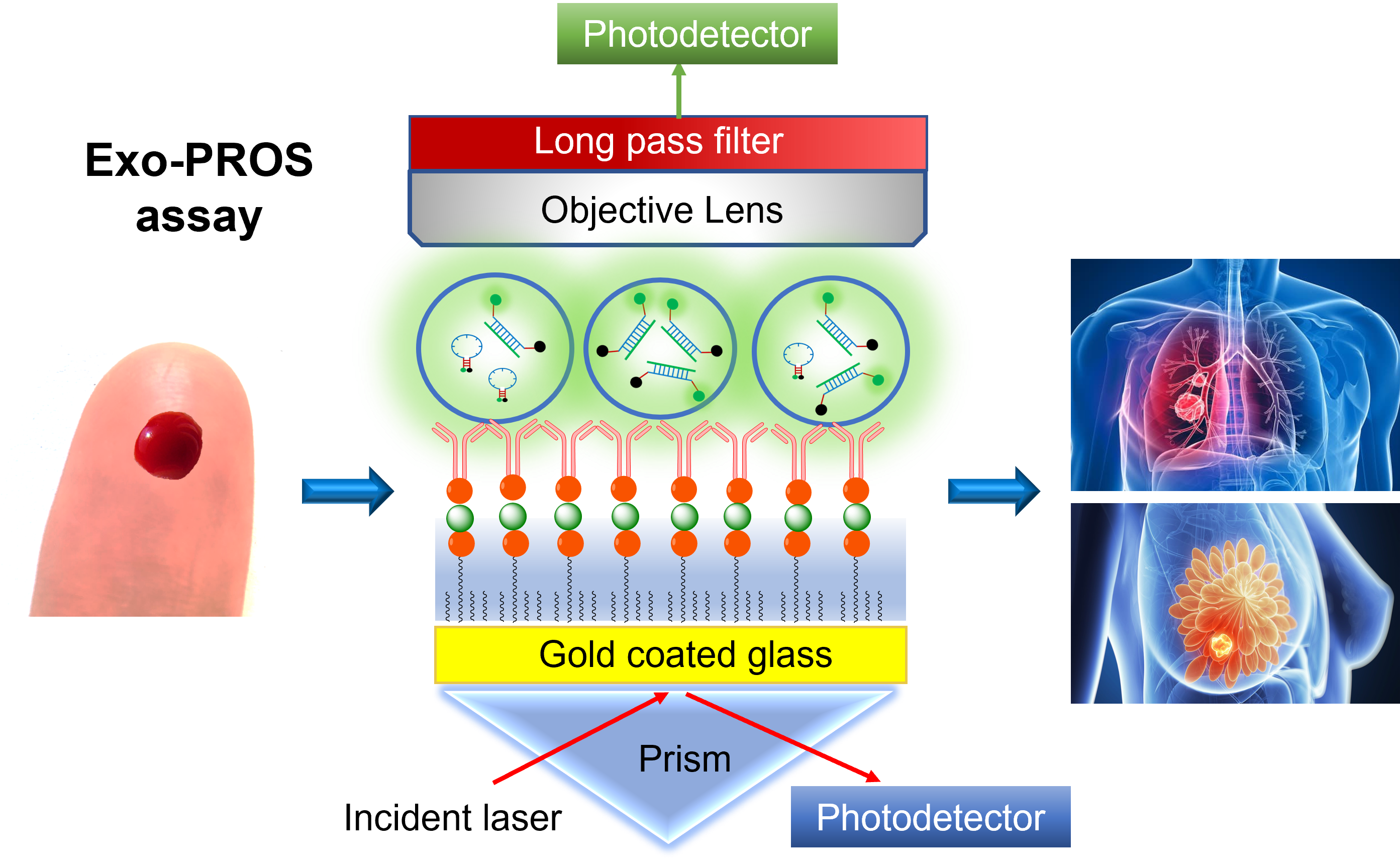
2.3. Exosome Protein RNA One Stop (Exo-PROS) biosensor simultanousely and in situ detects exosomal proteins and RNAs for cancer diagnosis
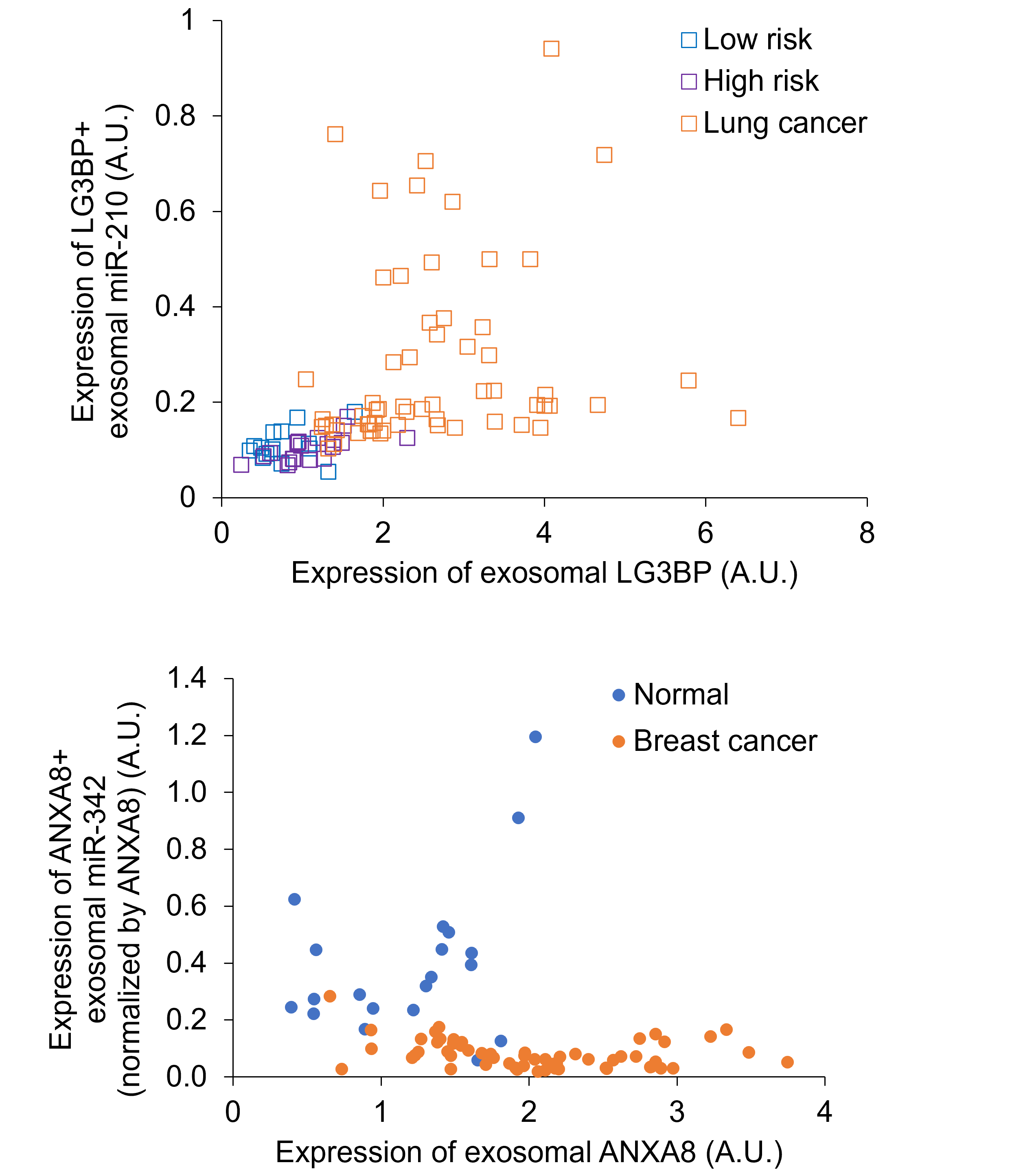
Tumor derived exosomes (TEXs) have emerged as promising biomarkers for cancer liquid biopsy. Conventional methods (such as ELISA and qRT-PCR) and emerging biosensing technologies mainly detect single type of exosomal biomarkers due to the distinct properties of different biomolecules. Sensitive detection of two different types of TEX biomarkers, i.e., protein and microRNA combined biomarkers may greatly improve cancer diagnostic accuracy. We developed an exosome protein microRNA one-stop (Exo-PROS) biosensor that not only selectively captured TEXs but also enabled in situ, simultaneous detection of TEX protein-microRNA pairs via surface plasmon resonance mechanism. Exo-PROS assay is a fast, reliable, low sample consumption and user-friendly test. With total 175 cancer patients and normal controls, we demonstrated that TEX protein-microRNA pairs measured by Exo-PROS assay detected lung cancer and breast cancer with 99% and 96% accuracy, respectively. Exo-PROS assay also showed superior diagnostic performance to conventional ELISA and qRT-PCR methods. Our results demonstrated that Exo-PROS assay is a potent liquid biopsy assay for cancer diagnosis.
.
.
Engineered Theranostics Laboratory