Study: Quick, pain-free breast imaging system shows promise in early clinical tests

Jul. 10, 2025
Lab News
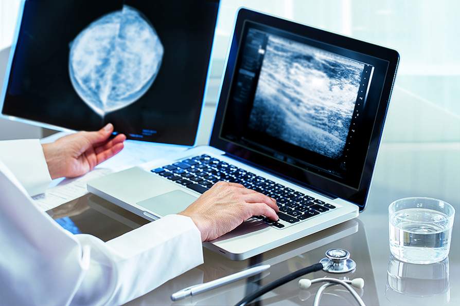
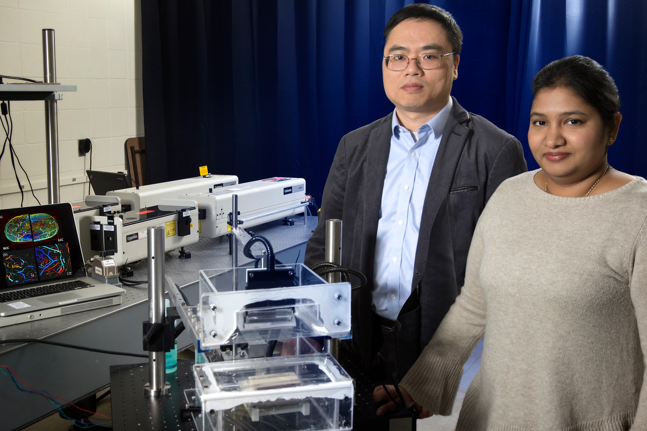
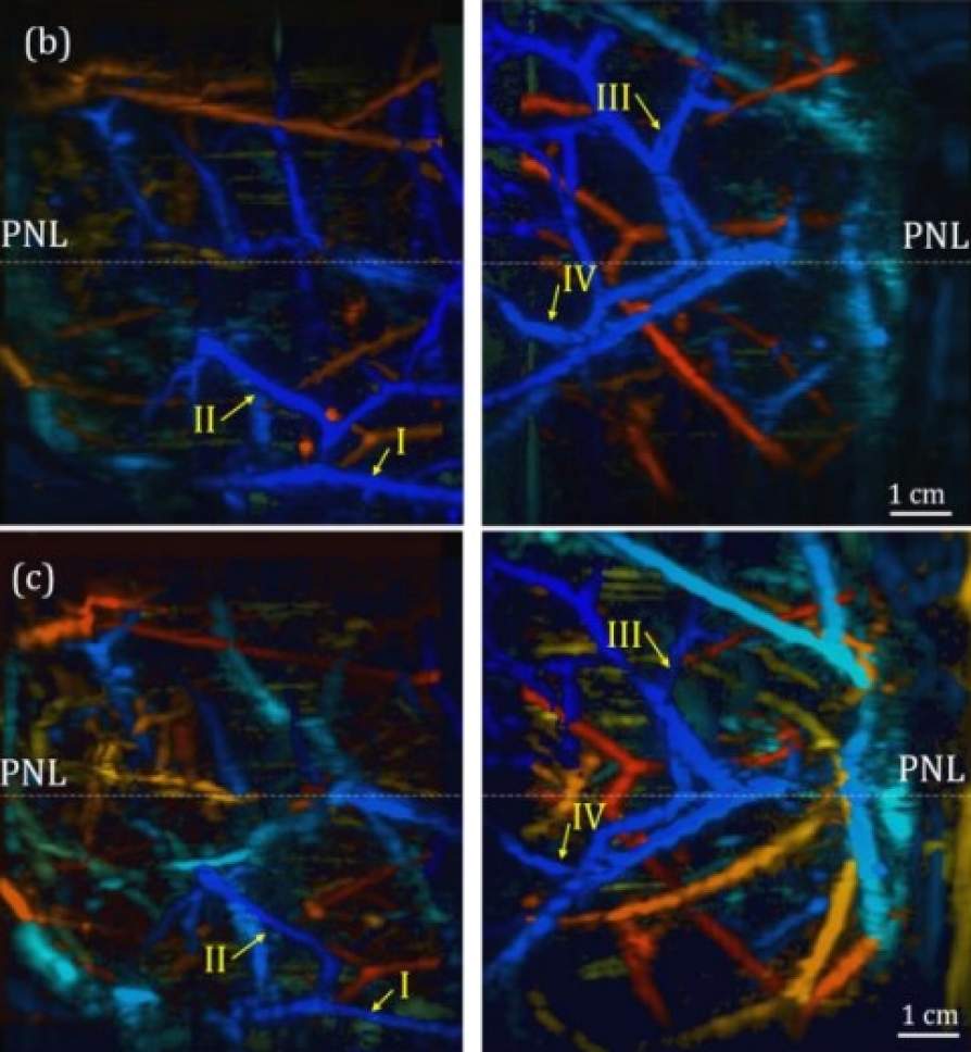
BUFFALO, N.Y. — A breast scan for detecting cancer takes less than a minute using an experimental system that combines photoacoustic and ultrasound imaging, according to a study in IEEE Transactions on Medical Imaging.