
 Research in the Pralle Lab at UB

The Biophysics of Cell Signaling in Immunology and Neuroscience.
Remote cell stimulation is a dream of neuroscientists because it would help tremendously in understanding how complex neuronal networks control behavior. Since 2005, optogenetics provides such a tool, but it requires visible light to penetrate to the neuron which often requires implanting optical fibers.
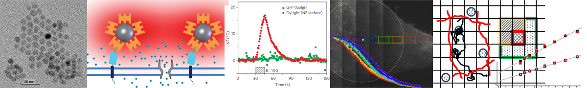
We have developed a remote simulation method based on radio-frequency magnetic field heating of super-paramagnetic nanoparticle of about 6-12 nm diameter, which are attached to or near to temperature sensitive TRPV1 ion-channels. We have demonstrated how this method can be used to remotely stimulate neurons in culture and in C. elegans [Nat.Nanotech. 5, 602-606]. Currently we are working with funding from HSFP and the NIH EUREKA program on improving the speed of the approach and to apply it to mammalian neuroscience in mice.
We also showed how fluorophores can be uses as molecular scale thermometers, and showed that we achieve membrane only heating. Currently, we investigate the local nanoparticle heating in further details. We have several postdoctoral positions available in this project. Please contact Dr. Arnd Pralle.
bimFCS and TNI resolve membrane ultra-structure
The cell membrane is a lipid bilayer, which hosts half of all proteins and conducts all cellular signaling with the environment. Initially it was assumed that membrane proteins diffuse freely without spatial order. Today, we know that cooperative activation of proteins often provides necessary signal-to-noise enhancement, and that certain cell signaling reactions require their components to be spatially segregated. Hence some spatial order is assumed to exist. One proposed clustering mechanism are Cholesterol-stabilized nano-domains enriched in certain lipids and proteins. However, these structures are transient and very small (10-50nm), which provides significant challenges to observing them in living cells. We have developed two methods to investigate these structures:Thermal noise imaging (TNI) use an optical trap to confine the motion of a tracer attached to a membrane protein to a 300nm x 300nm area of the membrane to increase statistics and the scattered laser light is used measure the position with high precision (us and nm). Using TNI we create high-resolution maps (4nm pixel size) of the cell membrane of three parameters (i) membrane bending stiffness, (ii) local diffusion of the tracked protein, and (ii) potential landscape transiently trapping the diffusing protein. We find nanodomains with increase stiffness, reduced diffusion and increase localized of raft proteins.
We have also developed a method to continuously monitor the interaction of fluorescently tagged membrane proteins with the nanodomains. The approach combines total internal fluorescence microscopy with fluorescence correlation spectroscopy (FCS) to record membrane protein diffusion on multiple length scales simultaneously. We refer to this method as binned-imaging FCS (bimFCS). Using bimFCS we have shown that cholesterol extraction, oxidization, or block of its synthesis by statin drugs all reduced the nanodomains. Using continuous monitoring on single cells, we have shown that dimerizing proteins, which transiently enter nanodomains, quickly increases the fraction of molecules associated with the nanodomains. Matching these experiments with Monte-Carlo simulations enabled us to quantitatively predict this increased domain association from the reduced dissociation probability of protein dimers from the domains.[ePub 2011, Physics ArXiv:1101.5087]
Immune cell membrane signaling
We use our methods (bimFCS,TNI) and others to investigate how the membrane ultra-structure modulates membrane signaling. We focus on two important effects in immune cell signaling:All mammals develop fever as a result of an infection despite the huge energy cost. Together with Dr. Elizabeth Repasky we study how moderate fever temperatures significantly increase the sensitivity of certain immune cells (T-cells) to stimulation.
All man-made vaccines contain adjuvants, which are empirically found substances increasing the immune response. Together with Dr. Terry Connell and funded by a NIH/NIAID grant we investigate the functional mechanism of a particular adjuvant, enterotoxin LTIIb.