Biophysics Modeling
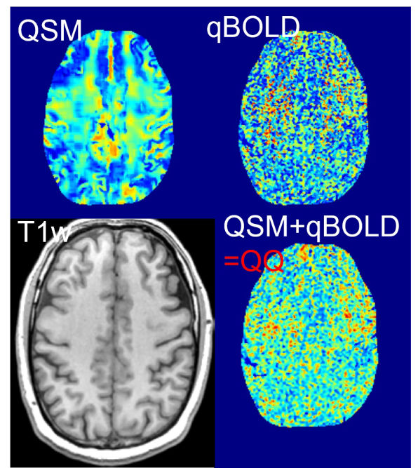
To obtain an accurate OEF map, we are developing realistic biophysics models based on MR physics. For instance, our recent model, namely QQ, considers deoxyhemogolobin effect, i.e. OEF effect, on MRI phase signal using
quantitative susceptiblity mapping (QSM) and magnitude signal using quantitative blood oxygen level dependent magnitude (qBOLD).
QQ can estimate OEF using a single routine MRI sequence without impractical vascular challenges unlike the other methods, which provides a high potential in clinical use.
See our recent work on an integrative OEF model, QSM+qBOLD=QQ
Data Processing
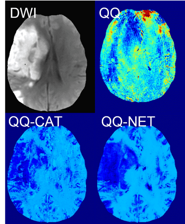
To solve biophysics models robustly, we are developing data processing algorithms including machine learning and deep learning approaches.
The biophysics models are complicated with having coupled, multiple model parameters.
Hence, they are difficult to solve, e.g., involved with poorly conditioned non-convex optimization.
To obtain reliable OEF, machine learning and deep leaerning algorithms have been developed.
For instance, cluster analysis of time evolution (CAT) is a machine learning algorithm that improves effective signal-to-noise ratio (SNR) substantially through clustering the voxels with similar signal patterns.
CAT enabled the detection of OEF abnormalities in stroke. Also, a deep learning approach, NET, led to improved OEF accuracy with substantionally faster, e.g., 150 times, reconstruction speed.
See our recent work on machine learning, CAT, CCTV, and deep learning, NET

Validation
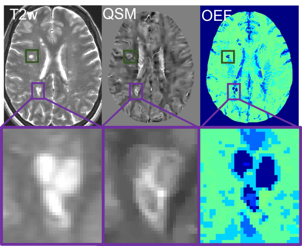
To validate our techniques, we are comparing ours with other imaging methods.
For clinical application, the validation is essential to investigate the accuracy of our techniques.
Our QQ provided consistent OEF values to a well-investigated MRI method, Calibrated fMRI, and the reference standard 15O-PET in healthy subjects.
See our recent work on comparison with Calibrated fMRI and 15O-PET
Clinical Application
To investigate disease progression and therapeutic strategies, we are applying our technique into neurologic disorders.
See our recent work in stroke, multiple sclerosis, dementia, brain cancer, pre-eclampsia
