Sample Research Projects
I.
System Matrix Modeling for
PET Reconstruction
To
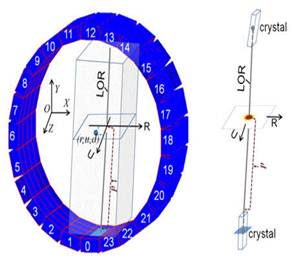
achieve optimal PET list-mode reconstruction, we develop a system matrix that
is based on the line-of-response probability density function (LOR-PDF). Each
LOR-PDF is a three-dimensional (3-D) function that describes fully the
distribution of LOR's contribution source. All the LOR-PDFs are sorted by the
LOR's incident angles to form a highly compact system matrix. The purpose of
this project is to achieve optimal statistical iterative PET image
reconstruction.
II. Animal SPECT Imaging on an Animal PET Scanner Animal
SPECT has become increasingly important in biomedical research. One major
limiting factor, however, for the wide acceptance of animal SPECT system is
its significant cost. Our group proposed a technology that would enable SPECT
imaging on an existing animal PET. This technology would allow the existing
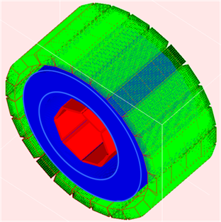
animal PET owners perform animal SPECT imaging Fig. 3.
A 3-D diagram illustrating the composition of the animal SPECT prototype
system. The detector ring of the animal PET is shown in green. The blue
annulus and red octagon are tungsten septa and knife edged lead plates which
are used to form multiple layers of fan-beam collimation for animal Fig. 4.
The bone image of a 25 gram Balb/C normal mouse
obtained with Tc-99m MDP tracer and the animal SPECT we developed.
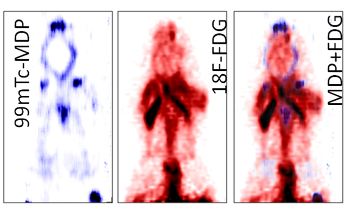
Fig. 5.
The PET and SPECT images of a mouse injected with mixed 99mTc-MDP (bone
agent) and 18F-FDG (metabolism) tracers obtained from the animal PET and
SPECT hybrid system. III.
Develop and support animal
PET applications
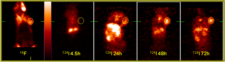
Fig. 6. An example result from project "Development of the See and Treat Multifunctional Photosensitizers" (in collaboration with Drs. Munawwar Sajjad and Ravindra K. Pandey). The 18F-FDG image on the left (coronal view) was acquired first as a reference after 90 minutes post-injection of 254 micro-Ci of activity to a tumor bearing C3H mouse. The mouse was then injected with 72 micro-Ci 124I- labeled derivative-of-Pyropheophorbide-a, a bi-functional (imaging and photo-dynamic therapy) agent. The mouse was imaged for 30 minutes at 4.5 hours, 24 hours, 48 hours and 72 hours post-injection. The tumor (yellow circle) uptake, as relative to the rest of the body, of the bi-functional agent increased over time, indicating promising perspective of therapeutic and monitoring application of the agent. The color palette (shown on the right of the 18F image) was scaled to the min/max of the transverse slice passing through the center of the tumor site (indicated by green-bars) in each dataset. The display scheme was same for all the images. Activity was injected via tail vein. |