Week 6
Modeling of small molecule-protein interactions
I. BASIC OPERATIONS OF SYBYL
The SYBYL package is optimized for operations with small molecules and their interactions with biopolymers. The program operation is similar to the InsightII and is menu-driven using an intuitive command structure. We will first practice some basic operations that allow you to manipulate biopolymers and their complexes.
Read in a molecule
File
Read
Select 1AQ7.pdb (This file contains the structure of an inhibitor bound to trypsin). (Read about this structure here)
Click OK
A dialog box appears asking "Center the molecule"
Click OK
The molecule is now displayed on the screen. You can control the size and orientation with the mouse buttons identically to how you do it in InsightII.
Use the "center-right" mouse buttons to fit the entire molecule to the screen size
We will now center the rotation axis around the inhibitor.
Build/Edit
Center
Click on the "Sets" button
In the new window, Click on the UNK_ATOMS
Then click OK in the two windows.
We will now 'undisplay' the water molecules.
View
Undisplay Atoms
Click on the "Sets" button
In the new window, Click on the Water
Then click OK in the two windows.
Now we will differentially color the enzyme and the inhibitor
View
Color
Atoms
Click on the "Sets" button
In the new window, Click on the UNK_ATOMS
Then click OK in the two windows.
A new window appears. Select a color (Red is good)
View
Color
Atoms
Click on the "Sets" button
In the new window, Click on the UNK_ATOMS
Click OK in this window.
In the selection window, click on the 'INVERT' Button
Click OK in this window.
A new window appears. Select a color (Green is good)
Now we will display the different parts of the enzyme active site: the residues responsible for substrate binding specificity and catalytic activity
View
Color
Atoms
Click on the "Substructures" button
In the new window, scroll down to Asp 189 and select it with your left mouse button
Click OK in this window.
Then click OK in the two windows.
A new window appears. Select a color (Yellow is good)
We have just highlighted the residue that recognizes the positively charged side chains in trypsin substrates and inhibitors. Now we will examine the "catalytic triad"-i.e., the residues that perform the actual bond cleavage in all serine protease.
View
Color
Atoms
Click on the "Substructures" button
In the new window, select His57, Asp102 and Ser195
Click OK in this window.
Then click OK in the two windows.
A new window appears. Select a color (White is good)
You can now use the mouse buttons to move the molecule and examine the interaction of the inhibitor with the enzyme-its binding site and why it functions as an inhibitor.
Now we will learn basic docking techniques. Before doing that we will have to clear the screen.
Build/Edit
Zap/Delete Molecule
Read in a molecule
File
Read
Select 1AQ7.pdb (This file contains the structure of an inhibitor bound to trypsin)
Click OK
A dialog box appears asking "Center the molecule"
Click OK
Center the rotation axis around the inhibitor.
Build/Edit
Center
Click on the "Sets" button
In the new window, Click on the UNK_ATOMS
Then click OK in the two windows.
In order to work with one molecule in the absence of another, the molecules must be in different work areas. The trypsin molecules and the inhibitor are stored in the same file in the database, so before we can dock the inhibitor, we have to extract it.
In order to extract a molecule, we have to have somewhere to put it. SYBYL has an option that allows you to divide the screen into several work areas, each of which can hold different molecules. We will start by dividing the screen into halves
Dividing the screen
On the left side of the SYBYL main window there is a series of buttons. The third one down looks like this: .
Click the button and then click the "Half" button, followed by the Q (quit) button.
Copy the cofactor to another molecule area
Build/Edit
Extract
Click on the "Sets" button
In the new window, Click on the UNK_ATOMS
Then click OK in the two windows.
A new window appears.
Select M2:<empty> as the molecule area
Click OK
Now we will delete the inhibitor from the original complex.
Build/Edit
Delete
Click on the "Sets" button
In the new window, Click on UNK_ATOMS.
Then click OK in the two windows.
Now we will name our inhibitor so that it will be easier to keep track of.
Build/Edit
Name molecule
Select M2
Click OK
Type inhibitor in the name block
Click OK
The docking process evaluates the suitability of an individual position/orientation by evaluating the interaction energies. As you already know, hydrogen bonds and charge interactions are important in molecular interactions. Thus, we must prepare the molecules to have the appropriate charges and numbers of hydrogens.
The first step in adding hydrogens is to ensure that SYBYL understands what kind of bonds are connecting the atoms in the molecule of interest.
Build/Edit
Modify
Bond
Select 'Type' as the element to modify.
Click OK
Select M2:inhibitor as the molecule
Then press the Bond Types button
In the next window, select uk (unknown
Click OK
Click OK in the Bond Expression window
The bond between the oxygen and the benzene moiety is highlighted in green, and a new window appears with a series of bond types 1 (=single), 2 (=double), etc.
Select 1 and click OK
The window disappears indicating that SYBYL understands all the other bond types in the molecule.
Now we will actually add the hydrogens.
Build/Edit
Add
Hydrogens
Select M2:inhibitor in the molecule area window
Click OK
Hydrogens are now added to the inhibitor.
Now we must assign the partial charges to the inhibitor atoms.
Compute
Charges
Gasteiger-Hò ckel
Select M2:inhibitor in the molecule area window
Click OK
A dialog box appears asking whether you want to change formal charges before computing charges
Select NO
Before beginning the docking process, color the molecule as we had done before.
View
Color
Atoms
Click on the "Sets" button
In the new window, Click on the UNK_ATOMS
Click OK in this window.
In the selection window, click on the 'INVERT' Button
Click OK in this window.
A new window appears. Select a color (Green is good)
View
Color
Atoms
Click on the "Substructures" button
In the new window, scroll down to Asp 189 and select it with your left mouse button
Click OK in this window.
Then click OK in the two windows.
A new window appears. Select a color (Yellow is good)
View
Color
Atoms
Click on the "Substructures" button
In the new window, select His57, Asp102 and Ser195
Click OK in this window.
Then click OK in the two windows.
A new window appears. Select a color (White is good)
There is a problem with the default settings of SYBYL. Before we can start the Docking procedure, we have to fix it.
Options
Tailor
Set
Select Dock in the Options window-Click OK
Select MAX_LATT_POINTS in the Tailor Option window-Click OK
A new window appears:
Change the value from 200000 to 400000-Click OK
Click END in the two Option windows
Now we are finally ready to begin docking!
To facilitate the docking, change the screen back to a single window:
Click the button and then click the "Full" button, followed by the Q (quit) button.
Compute
Dock
Select M2:inhibitor as the Ligand Molecule
Click OK
Select M1: as the Site Molecule
Click OK
Three new items appear on the screen.
Ordinarily, mouse movements move both the ligand and the target together. To have the mouse control only the ligand, press the F10 key on the keyboard
Now the translations and rotations only affect the ligand's movements.
Slowly move the ligand around and attempt to bring it into an energy minimum.
When you are done, exit back to the main menus by placing the cursor in the terminal window and pressing the ENTER key.
To return the appropriate mouse controls, click the button on the left row of buttons and select Global followed by the Q (quit) button.
To be sure the program behaves properly, we must 'reset' it. Do this by clicking the 'Reset' button on the left row of buttons and then click the 'Everything' button followed by the Q (quit) button.
Now we will learn how to build our own inhibitor and dock it into trypsin. Before doing that we will have to clear the screen.
Build/Edit
III. BUILDING YOUR OWN INHIBITOR
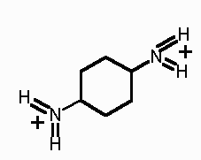
We will be building a benzene-based inhibitor of trypsin from benzene and arginine. (Read about the properties of this inhibitor here)
Build/Edit
Get Fragment
Scroll down and select Benzene from the option list
Click OK
Build/Edit
Get Fragment
Scroll down and select Arginine from the option list
Click OK
Select M2 as the molecule area
Click OK
We will now edit the arginine molecule to obtain the guanidine groups.
To move the arginine out where it can be operated on, click F10, and use the mouse to move the group out of the way.
To make life easier in terms of knowing which atoms to delete, we will label them.
View
Label
Atom name
Click on M2 in the Atom Expression window
Click the Atom Types button
In the next window, select C,O,N
Click OK
Click OK
Now we will create the guanidium group
Build/Edit
Delete
Bond
Click on the N2 nitrogen and the C5 carbon in the molecule display window.
Click OK
Now we will delete the "remainder" of the arginine residue.
Build/Edit
Delete
Substructure
Click on the Arg side chain in the molecule display window
Click OK
Now we will prepare for the 'chemical' joining of the benzene and the guanidium group.
Build/Edit
Delete
Atom
Click the proton on the N2 nitrogen in the molecule display window.
Click OK
Now join the benzene and guanidinium moieties.
Build/Edit
Other Tools
Join molecules
When the select Target window appears, click on one of the benzene protons
When the select Target window appears, click on the N2 nitrogen of the guanidinium group
Repeat this procedure placing the guanidinium group on the benzene at a para position with respect to the first guanidinium group.
The final molecule will look like this:
Now we will save this inhibitor
FILE
Save As
Select Benzene
Call the file inhibitor1
Click OK
Build/Edit
Zap/Delete Molecule
Now we will dock our inhibitor into the trypsin molecule.
File
Read
Select 1AQ7.pdb (This file contains the structure of an inhibitor bound to trypsin)
Click OK
A dialog box appears asking "Center the molecule"
Click OK
Build/Edit
Delete
Atom
Click on the "Sets" button
In the new window, Click on the Water
Then click OK in the two windows.
Now we will add H to the protein
Biopolymer
Add Hydrogens
Click on the All button
Click OK
In Option window (which hydrogens to add), select All
Click OK
Now we will load the protein's partial charges
Biopolymer
Load Charges
Click on the All button
Click OK
In Option window accept the Koll_Uni default
Click OK
To facilitate building of the new inhibitor complex, we will superimpose the new inhibitor on the old one and then delete the old one and begin the docking procedure.
View
Undisplay Atoms
Click the Sets button
Click OK
Click the Invert button
Click OK
Now read in the new inhibitor
File
Read
Select inhibitor1
Click OK
To help distinguish one from the other color the new inhibitor cyan
View
Color
Atoms
Click on M2
Click the ALL button
Click OK
Choose cyan and click OK
To help you in aligning the correct atoms, label the molecules
View
Label
Atom name
Click on M1 in the Atom Expression window
Click the Atom Types button
In the next window, select C,O,N
Click OK
Click OK
Repeat this procedure using M2 as the atom option.
Try using to align the one of guanidinium groups of the new inhibitor the guanidinium group o the old inhibitor. Use the F10 key to toggle mouse control between the new inhibitor and both molecules to aid this process.
Now we will re-display the protein.
View
Display Atoms
Click on M1
Click on the All button
Click OK
Now delete the old inhibitor
Build/Edit
Delete
Atom
Be sure M1 is selected
Click on Set
Select UNK_ATOMS
Click on OK in this and previous window.
Now Dock your inhibitor using the Docking procedure above.