Experiments in my laboratory currently focus on the developmental mechanisms that regulate nerve fiber outgrowth and the formation of nerve connections. One project utilizes invertebrate neurons whose large size and identifiable nature permit us to study the cellular mechanisms that regulate nerve growth. A second focus of my research involves the role of growth factors in the developing auditory and vestibular systems. Those experiments utilize chick embryos at various stages of development. Choose from the list below to learn more about these projects.
The unique characteristics of snail neurons provide many advantages for cellular studies of nerve growth. The large size of these neurons allow us to observe growth at the elongating tips (growth cones) of nerve fibers in great detail. Experimental manipulations can be performed on these structures with high precision. In addition, due to the smaller number of neurons in the snail nervous system and their simplified arrangement, we can identify individual neurons from one snail to another. These identified neurons are then plated, one at a time, into culture. Thus, the properties of a particular type of neuron can be determined without introducing the experimental variability that accompanies mixed populations of neurons.
Using snail neurons we have studied how electrical stimulation alters the movements of growth cones at the growing tips of cultured neurons. The generation of action potentials causes a decrease in the movement of growth cones as well as a change in their structure. These changes in growth cones are associated with alterations in internal calcium levels that we have studied using digital fluorescence imaging techniques:

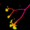
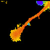
Spatial and temporal characteristics of calcium changes are important regulators of growth cone structure and function. Our recent data show that each type of neuron may have a specific level of calcium that induces a change in its growth cones. This implies that neurons may display a differential sensitivity to changes in calcium and thus to the stimuli that initiate these changes.
Another major interest is the role of the actin cytoskeleton in alterations in growth cone movements. Actin is a prominent and dynamic cytoskeletal component in the lamellipodia and filopodia of growth cones. Actin filaments are arranged as radially-aligned bundles that project into the filopodia and also as a more loosely organized meshwork that extends throughout the lamellipodium. These actin filaments exhibit a retrograde flow that moves filaments from the peripheral edge of the growth cone, where actin is assembled, to the central domain of the growth cone, where actin is disassembled. These characteristics are shared by other motile cells and provide a cellular basis for lamellipodial protrusion and motility. The large size of snail growth cones provide a unique opportunity to study the relationship between actin organization/dynamics and cell movement. We are studying actin organization and dynamics by injecting fluorescently labelled g-actin into neurons. This becomes incorporated into actin filaments in growth cones and is visualized by time-lapse imaging methods. Using this technique we are studing the role of actin filaments in living growth cones and how actin filaments may be influenced by molecules such as calcium during changes in growth cone function.
1. Williams, D.K. and Cohan, C.S. (1994) The role of conditioning factors in the formation of growth cones and neurites from the axon stump after axotomy, Dev. Brain. Res., 81:89-104.
2. Williams, D.K. and Cohan, C.S. (1995) Calcium transients in growth cones and axons of cultured Helisoma neurons in response to conditioning factors, J. Neurobiol. 27: 60-75.
3. Torreano, P. and Cohan, C.S. (1996) Electrically-induced changes in intracellular calcium in Helisoma neurons: regional and neuron-specific differences and implications for neurite outgrowth, J. Neurobiol. 32: 150-162.
4. Welnhofer, E., Zhao, L., and Cohan, C.S. (1997) Actin dynamics and organization during growth cone morphogenesis in Helisoma neurons, Cell Motil Cytoskel. 37: 54-71
The developing inner ear represents a complex sensory system in which the structures responsible for transducing sound and head position, the cochlea and vestibular apparatus, develop in close proximity to the peripheral sensory neurons that will innervate them. Under these conditions, the auditory and vestibular sensory neurons interact with their target sensory organs. This interaction involves the release of diffusible factors by the target organ, which influence the survival and outgrowth of the cochleovestibular neurons. Our experiments are directed at understanding the growth factor dependence of auditory and vestibular neurons throughout development. We have partially characterized diffusible factors that are released by the cochlea and vestibular organs early in development (embryonic days 4-6). Our experiments have characterized the biological effects of this factor and the time course over which it is released. Extensive comparisons with known growth factors indicate that it is not similar to any of the neurotrophins, CNTF, or other identified growth factors. At later developmental stages (E7-E16), other growth factors such as NT-3 and BDNF have significant effects on the cochleovestibular neurons. Our current experiments involve understanding how the growth factor requirements of auditory and vestibular neurons changes at different stages of embryonic development.
Visit the Center for Hearing and Deafness at the University.
1. Bianchi, L.M. and Cohan, C.S. (1991) Developmental regulation of a neurite-promoting factor influencing statoacoustic neurons, Dev. Brain Res. 64: 167-174.
2. Bianchi, L.M. and Cohan, C.S. (1993) Effects of neurotrophins and CNTF on developing statoacoustic neurons: Comparison with an otocyst-derived factor, Dev. Biol. 159: 353-365.
3. Hashino, E. and Cohan, C.S. Neurotrophic effects of BDNF and NT-3 on chick cochlear and vestibular ganglia in vitro, Assoc. Res. Otolaryng. (1996).
4. Hashino, E. and Cohan, C.S. CNTF prolongs BDNF-induced neurotrophic effects on chick vestibular ganglion neurons in vitro, Assoc. Res. Otolaryng. (1997).
Techniques currently in use in my lab include: low-light video and digital imaging microscopy, intracellular calcium imaging, time-lapse fluorescence imaging, intracellular and patch clamp electrophysiology, intracellular dye injection and immunohistochemical staining, immunoprecipitation, SDS PAGE, Western blot, ELISA, and cell and organ culture procedures.
This page maintained by C.Cohan
Last update: 5/10/97